


The Department of Cardiothoracic Surgery at Stanford University Medical Center takes pride in the rich tradition of excellence and pioneering firsts that have made it one of the top cardiac and thoracic programs in the nation. Our long and distinguished legacy of research dates back to the late 1950s — our most notable triumphs being the first adult human heart transplant in the United States, the world's first successful adult human combined heart-lung transplant, the first successful use of a ventricular device as a bridge to transplantation, the first thoracic aortic stent graft, and the development of the first integrated platform for minimally invasive heart surgery.
Our Department is comprised of three divisions:
internationally renowned for surgical leadership and expertise and a record of more than 30,000 cardiac procedures
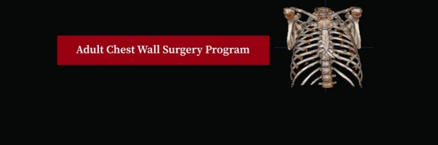
highly reputed for the management of patients with lung cancer, emphysema, esophageal cancer, and mediastinal diseases
one of the largest specialized pediatric cardiovascular surgery programs in the US, acclaimed for its contributions to improving survival from lethal cardiac malformations
Together, the Department of Cardiothoracic Surgery continues to improve patient health
through continual scientific innovation, revolutionary operative care, and exemplary surgical education.
Resident Applicants
For Medical Students
For Surgery Residents